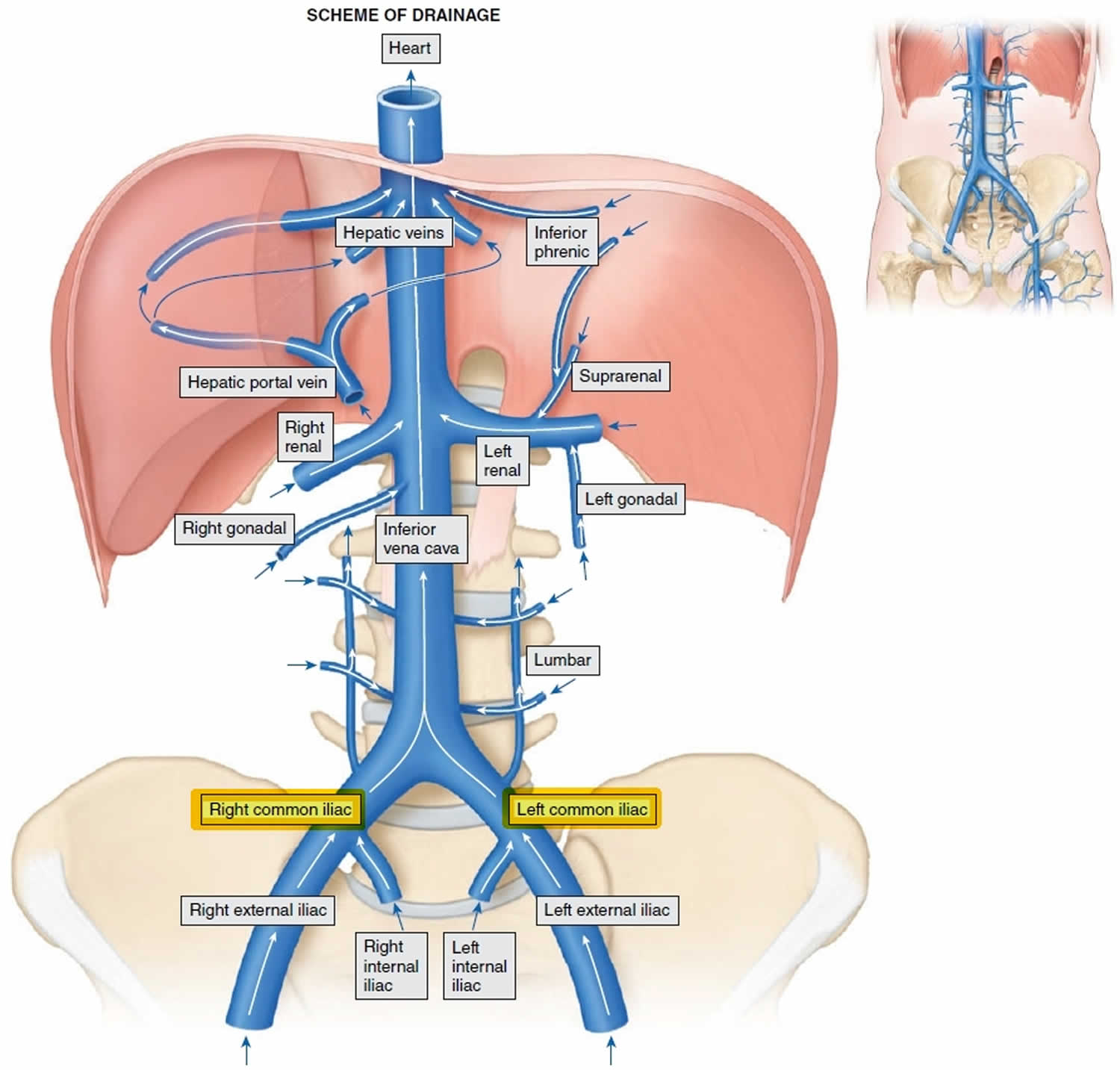
 Common iliac vein anatomy and function
Related Terms:Timothy R.S. Harward M.D., in , 201264 What is May-Thurner Syndrome? A proximal common left iliac vein narrowing or occlusion caused by an anatomical anomaly. Normally, the common left iliac vein passes under the right common iliac artery, as it joins the common right contralateral right vein to form the lower vena cava (IVC). The constant pulsation of the artery causes fibrosis of the underlying common left iliac vein, producing severe narrowing or total occlusion. When occlusion occurs, the entire left iliac venous system usually thrombos, and these patients are usually diagnosed with an iliofemoral DVT. Treatment with thrombolisis can dissolve the clot and discover the underlying pathology. If a wire can cross the narrowing, the area is treated with ball angioplasty and stent placement. In addition, these patients still require anticoagulation. Sara Paterson-Brown, in , 2010 The Venous System This is a low-pressure valve system to drain blood back to the heart. Flux fluctuates with the arterial pulse while muscle pumps further promote the flow in the extremities and the inspiration increases the flow in the lower and upper vena cava (IVC and SVC) centrally. Except the portal's circulation, the veins generally follow the pattern and path of the arteries and have a sympathetic interiorization. The lower vena cava Common iliac veins are joined to form the IVC (Fig. 5.5) behind the right outer artery in L5. The IVC ascends through the abdomen to the right of the aorta by piercing the central tendon of the diaphragm in T8. Receive: Segmental lumbar veins The right gonadal vein (the left gonadal vein is drained into the left renal vein) The renal and suprarenal veins The liver veins The inferior ferrine veins. Colateral Venous Drainage Ways There is an extensive network of possible collateral circulations that open when the IVC thrombosis occurs. Superficial venous canals that may eventually drain the upper vena cava are:EpigastricCircumflex iliac Fantastic epigastric and lateral chest (via toracoepigastricate vein)Internal chestPosterior intercostal External PosteriorLumbovertebral. The deep channels that provide deep anastomosis are:AzygousHemiazygousLumbar. The vertebral venous plexus also provides effective collateral circulation between IVC and SVC. This collateral circulation is so efficient that even when there is substantial obstruction to the venous flow by a large deep vein thrombosis in the illic vessels, there may be a lack of symptoms or clinical signs. The portal venous drainage and the anastomous venous porosis The venous portal drains blood to the liver of the abdominal part of the digestive canal (except the anus), spleen, pancreas and bladder. The upper and lower mesenteric veins join the splenic vein behind the pancreas to form the portal vein that carries blood to the liver, which in turn is drained by the hepatic veins that pass to the IVC. This pathway can be obstructed by causing portal hypertension and then open collateral between the portal and systemic venous systems: Lower esophagus – influents of: left gastric with hemiacigous/azygous Anal rectal edge with mid and lower rectal area Caput medusa – influent of the left branch of the portal vein (paraumbilical) Vertebral Column Vegetable drainage of both the internal and external drainage of the vertebral plexo to regional segmental veins that provide potential communication with systems that also drain segmented. This is a system largely without valves and therefore the propagation of malignancy is possible (especially probably from the breast, uterus, prostate and thyroid): Pelvic viscera through the sacral side vessels Abdomen via the lumbar veins The breast through the subsequent intercostals Neck through the vertebral vein. S. Jacob MBBS MS (Anatomy), in , 2008 In addition to the two common illogical veins, the lumbar veins, the right gonadal veins, the kidney veins, the right suprarenal vein, the right lower fenica vein and the three hepatic veins and several hepatic accessory veins drain into the vena cava. The left gonadal veins, the left adrenal vein and the lower left ferina vein are tributaries of the left renal vein. Priscila Gisselle Sanchez Aguirre, ... José I. Almeida, in , 2019 In contrast to the right common iliac vein, which almost vertically ascends to the lower vena cava (IVC), the left common iliac vein takes a more transversal course under the right common iliac artery, which can compress it against the lumbar column before entering the vena cava. This compression causes the stasis of the blood, which is an element of the Virchow triad that can precipitate the deep vein thrombosis (DVT). In about 40% of patients, the precursor of iliac venous obstruction is not thrombosis, but rather a non-trombotic iliac lesion (NIVL). Also known as May-Thurner1 syndrome or iliac vein compression syndrome,2 is caused by web formation3 and obstruction. A non-trombotic block is defined as a clinical record absent from DVT along with missing findings on imaging, such as contrast and ultrasound venography. This lesion is classically found in the common left vest vein of younger females; but it is not uncommon in men or in older patients, and may involve the right member. At least 15 per cent of the extremities with primary disease have proven to have common and external iliac vein stenosis. 4 As described in the Fig. 18.1, the right common ilic artery always crosses the left common iliac vein in the confluence of the IVC and is where the classical proximal NIVL is produced. In 75% (majority model), the right iliac artery continues its course to cross close to the external iliac vein level, in which case it can induce the right distal NIVL. In the minority pattern (22%), the right iliac artery, common crosses the right iliac vein, common, and then descends on a longer length of the outer iliac vein and can thus induce a proximal or distal right NIVL. The left distal lesion may be related to the cross of the vein by the left hypogastric artery. These anatomical variations can explain why proximal NIVLs occur more often on the left side than on the right side, why the left injury is focal and the right is less so, and why the distal NIVL occurs equally on both sides. 5Although traditional venous corrective surgery concentrated on surface and deep venous reflux correction under the inguinal ligament, the introduction of minimally invasive venous stent using venography and intravascular ultrasonography (IVUS) provides the ability to treat the "obstructive" component of the disease above the inguinal ligament. The emphasis on the IVUS as a key diagnostic tool was promulgated by Raju and Neglén who have shown that the vialous stent is only sufficient to control symptoms in most patients with combined output obstruction and deep reflux. 6Timothy K. Williams, Julie Ann Freischlag, in , 2013Pathophysiology The right CIA crosses before the left IVC in the confluence with the aorta and the vena cava. The mechanical compression of the left IVC can occur, with the consequent lower-intensity venous hypertension and thrombosis. However, the mechanical compression of the left IVC is often identified in patients who are asymptomatic, thus putting in question the contribution of mechanical compression in the syndrome. 70.71 Protrombotic states have been involved in the appearance of this syndrome,72–74 and it is not known whether mechanical compression is the only instigator of symptoms or whether other factors are involved for most patients. Luis M. Chiva, Javier Magrina, in , 2018 The IVC receives the venous flow of the right and common illogical veins and is located on the right of the aortic fork. Similar to the arterial correspondents, the outer iliac vein drains mainly the lower extremities, while the inner iliac vein drains the pelvic viscera, the walls, the gluteal region and the perineo. In most cases the main veins are mirror images of their arterial counterparts. However, smaller vessels may vary from one individual to another. The lower epigastric veins, deep and pubic iliacs are all the pelvic affluents of the outer ilic vein. The outer iliac vein is the superior continuation of the femoral vein. The nomenclature of the vessel changes in the middle-in-in-uinal point, after the inguinal ligament. The deep iliac vein crosses the anterior surface of the outer iliac artery before entering the outer iliac vein. Inferior to the entry point of the deep iliac vein, the lower epigastric vein enters the outer iliac vein cefalad to the inguinal ligament. The pubic vein forms a bridge between the shutting vein and the external ilic vein. On the left side, the outer iliac vein is always medial to its corresponding artery. However, on the right side, it begins in a medial position and gradually becomes later, as it approaches the melting point. The inner iliac vein receives as tributaries the medium rectal veins, shutters, lateral sacral, lower gluteals and higher gluteals. The shutter vein enters the pelvis through the shutter foramen, where it takes a posterosuperior route along the lateral pelvic wall, deep into its artery. In some cases the container is replaced by an expanded pubic vein, which then binds to the external ilic vein. The upper and lower gluteal veins accompany the veins of their corresponding arteries. The tributaries of the upper gluteal veins are named after the branches of the corresponding artery. They pass over the piriformis and enter the pelvis through the larger cytic foramen before joining the inner iliac vein as a single branch. The lower gluteal veins form anastomous with the first piercing vein and the circumflex medial femoral vein before entering the pelvis through the larger cytic foramen. The medium rectal vein is a product of the rectal venous plexo that drains the mesorectum and the rectum. It also receives affluents from the bladder, as well as specific genus affluents from the prostate and seminal vesicle or the posterior wall of the vagina. It ends in the inner iliac vein after traveling along the pelvic part of the ani levator. Finally, the sacral side veins travel with their arteries before entering the inner ilic vein. The internal and external illogical veins are joined in the sacroiliac joint, on the right side of the fifth lumbar vertebrae, to form the common ilic vein. The right common ilic vein is almost vertical and shorter than the left common ilic vein, which takes a more oblique course. The right-brain nerve crosses the right-hand common iliac vein later; the sigmoid vessels mesocolon and upper rectangles cross the common left vein earlier. The internal poddal vein is drained into the inner iliac vein, while the medium sacral veins are drained directly into the common ilic vessels. The medium sacral veins are joined in a single glass before entering the common left iliac vein. The internal cannon veins receive lower rectal veins and clitoris and labial or penis bulb and scrotal veins before joining the common ilic vein (Figs. 2,27 and 2.28). Tim Hartshorne, in , 2011 Anatomy and Flow Guidelines The IVC is formed by the confluence of the common left and right iliac veins around the level of the fifth lumbar vertebra. It is located on the right of the aorta and prior to the right aspect of the spine. In its upper abdominal segment it runs in a groove in the posterior aspect of the liver and is separated from the aorta by the right crus of the diaphragm before drilling the diaphragm at the level of the eighth thoracic vertebrae. Anatomical variations occur but many are rare. 79,80 The most common variations occur below the level of the renal veins and involve the transposition or duplication of the IVC, resulting in a left-sided vena vena cava or lower double vena cava where the common iliac veins do not unite but continue better as paired vessels with the left component that join the right side at the level of the left renal vein (Fig. 40.32). The size of the IVC can change significantly in response to a variety of factors described in table 40.6. In particular, changes in diameter with breathing can be observed due to changes in intrathoracic and abdominal pressure. During inspiration, the venous return is increased as the blood is suctioned in the chest by negative intrathoracic pressure and the abdominal cava is compressed by increased intraabdominal pressure resulting from the drop of the diaphragm; therefore, the diameter of the IVC decreases during inspiration. On the contrary, during the expiration of the intratoraic pressure increases, which reduces blood flow in the chest, and the intraabdominal pressure decreases as the diaphragm rises, which results in the increase in the diameter of the cavalry. In addition, changes in the right-hand head pressure to normal heart disease or activity will affect the flow and size of the lower vena cava. Flow patterns recorded in the IVC are usually phases with breathing and with a superimposed pulpit due to the reflected right atrial pressure waves (Fig. 40.33). These characteristics are generally more pronounced in the proximal IVC. Changes in caval diameter variation have been correlated with blood loss81 and fluid dehydration/overload status in patients with trauma and renal replacement therapy. 82GEZA MOZES, PETER GLOVICZKI, in , 2007ANATOMY OF THE ABDOMINAL AND PELVIC VEINS The lower vena cava (IVC) begins in the confluence of the common iliac veins and ascends on the right side of the spine, passes through the tendentious portion of the diaphragm, and after a short course (approximately 2.5 cm) in the chest ends in the right atrium at the level of T9. In the upper abdomen the IVC is found after the duodenum, the head and neck of the pancreas, the lower sac and the liver. The IVC intrahepatic portion is found in a slot along the back aspect of the enclosed lobe. The IVC tributaries are the paired lumbar and renal veins and the liver veins, and on the right side the right, suprarenal and lower gonadal veins are also drained into the IVC (see Figure 2.4). The left adrenal veins and gonadal veins are joined to the left renal vein, the lower left ferina vein is drained into the left adrenal vein. In case of IVC obstruction, communication between the veins of the chest and abdominal wall (torocoepigastrica, internal thoracic and epigastric veins), lumbar anastomosis and vertebral plexies provide important collateral pathways. Common iliac veins begin in the sacroiliac joint on both sides and end in L5, where they form the IVC. The only tributary of the right common iliac vein is the right ascending lumbar vein; the common left iliac vein drains the left ascending lumbar veins and median sacral (see Figure 2.4). The right common iliac vein is deferred-lateral to the right common iliac artery. The distal segment of the common left iliac vein is medial and post to the common left iliac artery, the proximal segment is after the right iliac artery and distal aorta. Compression of the proximal common left iliac vein may occur due to excessively uplifting arterial structures. The outer iliac vein begins at the level of the inguinal ligament, extends along the pelvic edge and ends before the sacroiliac joint where the external and internal ililiac veins form the common iliac vein. On the right the distal external iliac vein is medial to the artery; however, as it ascends, more likely, it extends after it. The left outer iliac vein remains median to the artery throughout its course. The tributaries of the outer vein are the lower epigastric veins, deep and pubic circumflex iliacs. The internal iliac vein is a poster-medial to the internal iliac artery on both sides. The short trunk of the internal iliac vein is formed by the confluence of the extra and intrapelvic venous tributaries. Extrapelvic affluents include the gluteal veins (superior and lower), internal throttle and shutter, which drain the pelvic wall and perineum. The intrapelvic affluents of the inner iliac vein are the sacral and visceral lateral veins (quare rectal, bladder, uterina and vaginal), which drain the venous plexus of precral and visceral pelvic (rectal, vesical, prostatic, uterine and vaginal). Both the IVC and the common iliac veins are invalid. There is usually a valve in the outer iliac vein, however, it is often without any valve. Giovanni Aletti... Vanna Zanagnolo, in , 2018Step 3: Elimination of the Common Iliac Nodes The dissection of the right and left sides is different. On the right side, the common iliac vein is lateral to the artery, while on the left side the vein is below and medial to the artery. The lateral margins and means of dissection are identified; the areolar tissue is incised for the length of the vessels and is dissected with the use of vena retractors as necessary. Common iliac veins may have important tributaries including the iliolumbar vein and, on the left side, the middle sacral vein. The common lateral lymph nodes are typically eliminated as a component of the complete dissection of the pelvic node. R. Douglas Orr, in , 2019L5-S1 Level Exposition To expose the L5-S1, work in the interval only medial to the left common iliac vein. Palp the disk. Place the right side retractor blade and a top holding the aortic fork. Use a comet dissector to spread the tissue rotundamente out of the annulus. Find the middle-line sacral vessels. There are usually two veins and an artery, but, like with the iliolumbar vein, the vessels can be very variable. 16 If the vessels are small, they can be taken with bipolar and divided captivity. The larger veins, up to 1 cm, can be found and must be tied. Often working on the bed after the divided vessels provides the easiest way to remove the tissue from the album annulus. The left common iliac vein often overlaps the superolateral aspect of the disk and must be bluntly swept from the disk and maintained under a retractor sheet (Fig. 6.9). Recommended Publications: We use cookies to help provide and improve our personalized service and content and ads. By continuing to accept . Copyright © 2021 Elsevier B.V. or its licensors or collaborators. Direct Science ® is a registered trademark of Elsevier B.V.ScienceDirect ® is a registered trademark of Elsevier B.V.
Server Error Please try later. Accessibility Links Search modes Search resultsVena Iliac - WikipediaVena ilíaca interna - WikipediaVena iliaca common - WikipediaVena iliaca common: Anatomy and drainage ANTE KenhubVena iliac common ¦Radiología Reference article ...Pelvic veins, lymphatics and nerves: Anatomy and drainage ...External ilíacical view - Wikipedia External, internal and common illogical veins - YouTubeFooter links

The Pelvic Veins - External - Internal - Common Iliac - TeachMeAnatomy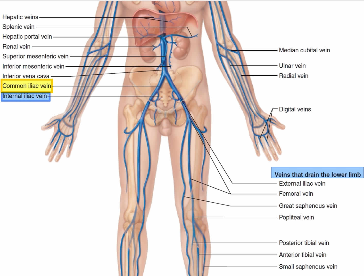
Common iliac vein anatomy and function
iliac arteries
External iliac vein - Wikipedia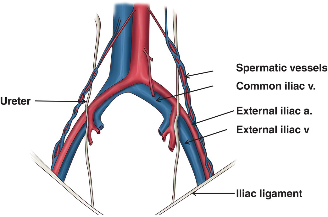
Iliac injuries (Chapter 29) - Atlas of Surgical Techniques in Trauma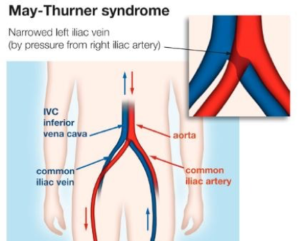
Balloon angioplasty and stenting for obstructive venous lesions like May-Thurner - Vascular Care Centre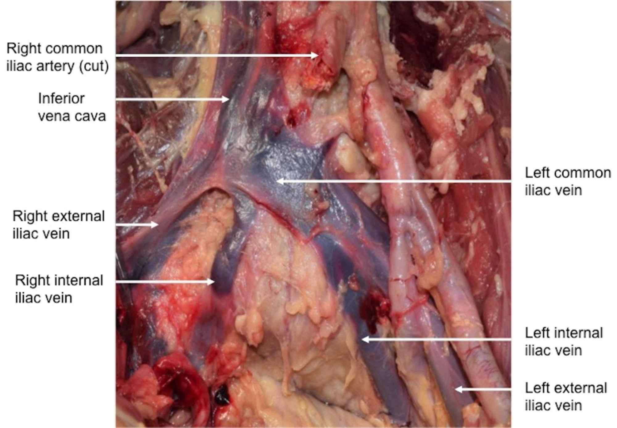
Cureus | Absence of the Right Common Iliac Vein with the Right Internal Iliac Vein Arising from the Left Common Iliac Vein: Case Report
Iliac Anatomy - Anatomy Drawing Diagram:background_color(FFFFFF):format(jpeg)/images/library/7837/985_RectumBloodSupply.png)
Iliac vein compression syndrome - Case with images | Kenhub
May Thurner Syndrome | Iliac Vein Compression Diagnosis & Treatment:format(jpeg)/images/article/en/iliac-vein/IZTVpFAkcNyN2f9VNoLXQ_Q2O5c1yqDSGOVBST5C8XKg_V._iliaca_communis_m01.png)
Common iliac vein: Anatomy and drainage | Kenhub
Solved: PAL: Models > Cardiovascular System - Veins > Quiz... | Chegg.com
Common Iliac Vein - an overview | ScienceDirect Topics
Circulatory Routes | Boundless Anatomy and Physiology
Iliac vein | anatomy | Britannica
Chapter 32 – Iliac Vessel Injuries | Anesthesia Key
Lumbar veins - Wikipedia
ObjectivesObjectives At the end of the lecture, the student should be able to: Define the 'vein' and understand the general principle of the veins. Define. - ppt download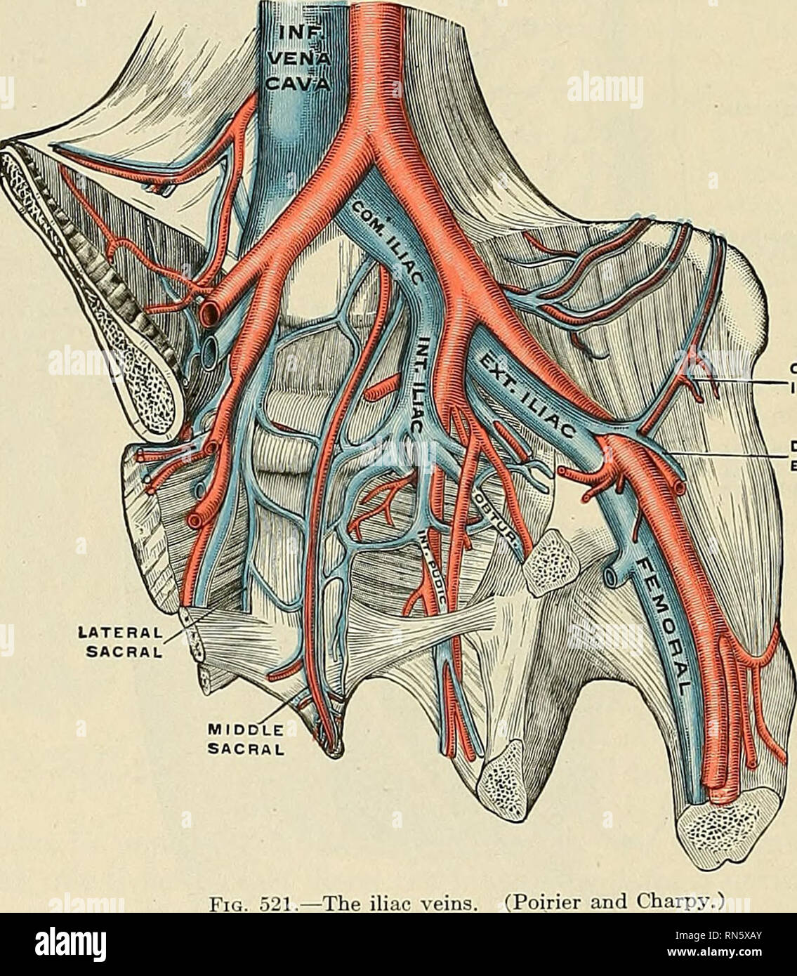
Internal Iliac Vein High Resolution Stock Photography and Images - Alamy
Circulatory Routes | Boundless Anatomy and Physiology
Iliac vein | anatomy | Britannica
THE VEINS Introduction The Pulmonary Veins The Systematic Veins. - ppt video online download
Common Iliac Artery - an overview | ScienceDirect Topics
Solved: PAL: Models > Cardiovascular System - Veins > Quiz... | Chegg.com/the-blood-supply-of-the-pelvis-87313859-548b47527f2547d9bff9040f0bb1db4e.jpg)
Common Iliac Artery: Anatomy, Function, and Significance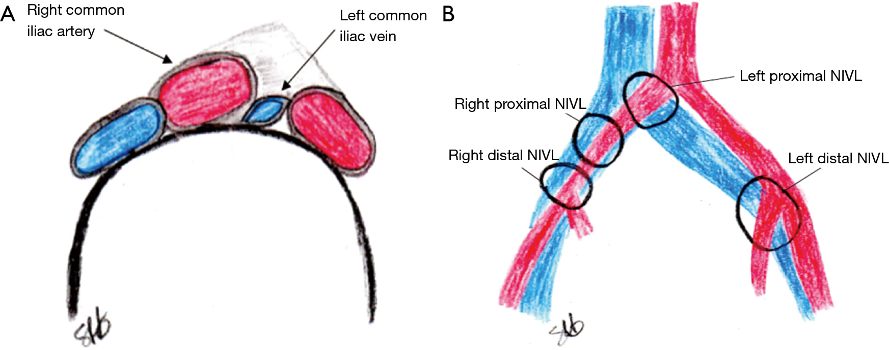
Re-intervention for occluded iliac vein stents - Black - Cardiovascular Diagnosis and Therapy![Solved] I am having trouble labeling this. I have looked through so many pictures, but can find one that comes close. Can you help? | Course Hero Solved] I am having trouble labeling this. I have looked through so many pictures, but can find one that comes close. Can you help? | Course Hero](https://www.coursehero.com/qa/attachment/4303959/)
Solved] I am having trouble labeling this. I have looked through so many pictures, but can find one that comes close. Can you help? | Course Hero
Ultrasound image of the veins of pelvis – the left common iliac vein... | Download Scientific Diagram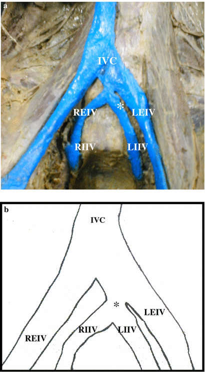
Rare variation in course and affluence of internal iliac vein due to its anatomical and surgical significance | SpringerLink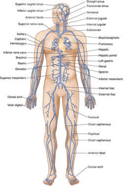
Deep circumflex iliac vein | definition of deep circumflex iliac vein by Medical dictionary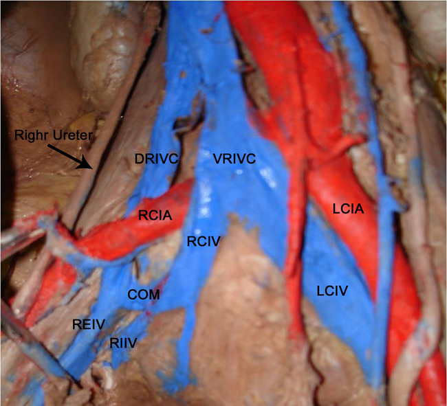
Article Fulle Text
Common Iliac Vein Anatomy, Function, and Diagram | Body Maps
The abdominal aorta bifurcates at the level of the fourth lumbar vertebra to form the two common iliac arteries, each… | Arteries anatomy, Arteries, Abdominal aorta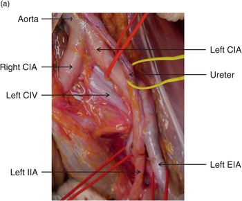
Iliac injuries (Chapter 29) - Atlas of Surgical Techniques in Trauma
a Right internal iliac vein has a course that stretches cranially (+),... | Download Scientific Diagram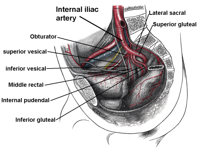
Branches of Internal Iliac Artery | The Lecturio Medical Online Library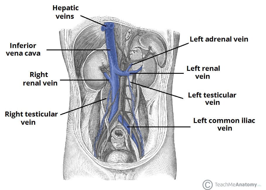
Venous Drainage of the Abdomen - TeachMeAnatomy:background_color(FFFFFF):format(jpeg)/images/library/12546/Iliac_and_femoral_nerves_and_vessels.png)
Iliac vein compression syndrome - Case with images | Kenhub
Abdominal cavity - AMBOSS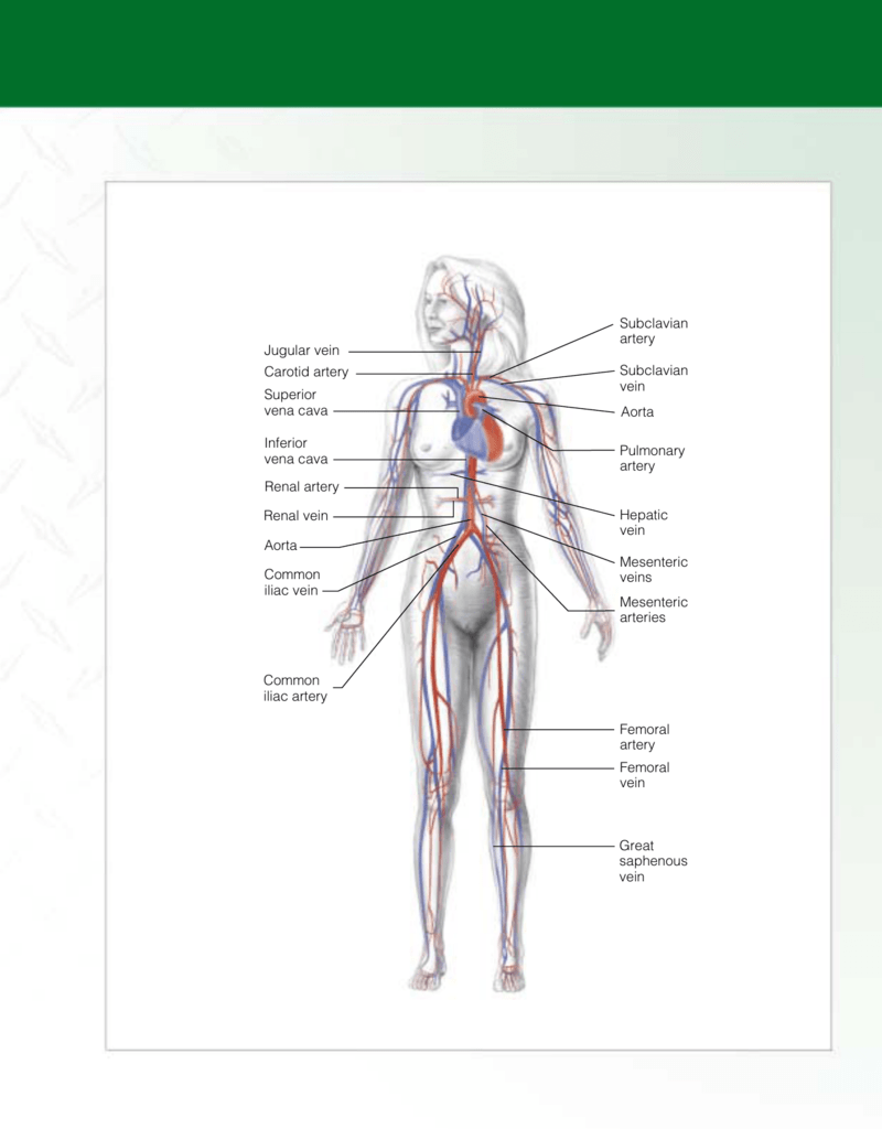
Jones-PA&P-ch01 #3 - Paramedic EMS Zone
 Common iliac vein anatomy and function


:background_color(FFFFFF):format(jpeg)/images/library/7837/985_RectumBloodSupply.png)

:format(jpeg)/images/article/en/iliac-vein/IZTVpFAkcNyN2f9VNoLXQ_Q2O5c1yqDSGOVBST5C8XKg_V._iliaca_communis_m01.png)


/the-blood-supply-of-the-pelvis-87313859-548b47527f2547d9bff9040f0bb1db4e.jpg)


:background_color(FFFFFF):format(jpeg)/images/library/12546/Iliac_and_femoral_nerves_and_vessels.png)


Posting Komentar untuk "the two common iliac veins form the"